

Optimum LIFT™ Mini Lift : Targeted Lower Face & Neck Correction
26 janvier 2026
Jaw Asymmetry Surgery : The surgical approach to facial harmony
2 février 2026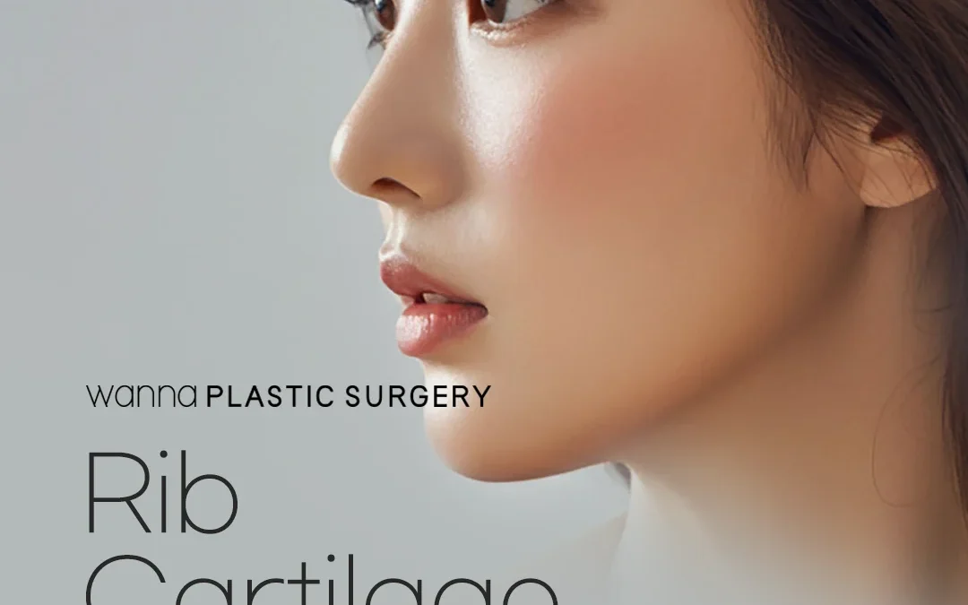
Unveiling the New You : Life after Rib Cartilage Rhinoplasty
Rib cartilage rhinoplasty represents a sophisticated and often indispensable technique in the reconstructive and aesthetic armamentarium of facial plastic surgeons. It is a procedure that leverages the body’s own robust and plentiful cartilage from the rib cage to reshape and augment the nose, addressing a spectrum of complex nasal deformities and aesthetic concerns.
While septal and ear cartilage are often the primary choices for rhinoplasty due to their proximity and ease of harvest, situations arise where these sources are insufficient or unsuitable, propelling rib cartilage to the forefront as the preferred grafting material.
Rib cartilage rhinoplasty is a powerful and transformative surgical procedure, offering solutions for some of the most challenging nasal deformities and aesthetic goals. It stands as an indispensable tool in the reconstructive surgeon’s repertoire, particularly when other autologous cartilage sources are insufficient. While it presents unique challenges, primarily related to donor site morbidity and the inherent warping potential of the cartilage, meticulous pre-operative planning, precise surgical technique, and diligent post-operative care can lead to highly successful and durable outcomes.
What are the advantages of rib cartilage rhinoplasty?
Rhinoplasty, commonly known as a « nose job, » is a surgical procedure designed to reshape the nose, improving its proportion and overall harmony with other facial features. It can correct breathing problems associated with nasal structure and address congenital defects, trauma, or previous unsuccessful surgeries. The nose, a complex three-dimensional structure, is composed of bone, cartilage, and soft tissues, all contributing to its form and function.
At the core of successful rhinoplasty lies the meticulous manipulation and structural support provided by cartilage grafts. These grafts are crucial for augmenting deficient areas, providing structural support to prevent collapse, refining the nasal tip, straightening the dorsum, and improving airway patency. The ideal grafting material should be biocompatible, resistant to infection and resorption, easily carved, and provide stable, long-lasting results.
Historically, various materials have been used, including synthetic implants (e.g., silicone, Gore-Tex) and allografts (cadaveric cartilage). However, autologous cartilage—cartilage harvested from the patient’s own body—remains the gold standard due to its superior biocompatibility, minimal risk of rejection or infection, and long-term stability. The primary autologous sources include:
- Septal Cartilage: Located within the nasal septum, it is often the first choice due to its proximity, ease of harvest, and ideal stiffness. However, its supply is limited, especially in revision cases or noses with pre-existing septal deviations or perforations.
- Auricular (Ear) Cartilage: Harvested from the concha of the ear, it is more pliable and curved, making it suitable for tip refinement, alar grafts, or camouflaging minor dorsal irregularities. Its supply is also limited, and its flexibility makes it less ideal for strong structural support.
- Costal (Rib) Cartilage: This is the focus of our discussion. Rib cartilage offers an abundant supply of strong, straight cartilage, making it invaluable for major structural reconstruction and augmentation.
Why Rib Cartilage?
Rib cartilage emerges as a critical resource when septal and ear cartilage are depleted or insufficient for the reconstructive goals. Its key advantages include:
- Abundant Supply: A single rib can provide a substantial amount of cartilage, enough for extensive nasal reconstruction.
- Structural Strength: Rib cartilage is robust, offering excellent support for dorsal augmentation, columellar struts, and caudal extension grafts, which are critical for establishing nasal projection and length.
- Versatility: It can be carved into various shapes and sizes, from thin sheets to substantial blocks, accommodating diverse reconstructive needs.
- Autologous Nature: Being the patient’s own tissue, it integrates well, minimizing the risk of foreign body reactions, infection, and extrusion compared to alloplastic implants.
However, rib cartilage also presents unique challenges, primarily related to donor site morbidity (pain, scar, potential for pneumothorax) and the inherent tendency of cartilage to warp or bend over time, a phenomenon that requires specialized carving techniques to mitigate.
Indications for Rib Cartilage Rhinoplasty
Rib cartilage rhinoplasty is typically reserved for complex cases where significant structural support or augmentation is required, and other cartilage sources are either unavailable or inadequate. The primary indications include:
Revision Rhinoplasty
This is perhaps the most common indication. Patients undergoing revision rhinoplasty often present with depleted or damaged septal and ear cartilage from previous surgeries. Common issues in revision cases include:
- Saddle Nose Deformity: A collapse of the nasal dorsum, often due to aggressive septal cartilage resection or trauma, creating a « saddle-like » depression. Rib cartilage is ideal for rebuilding the dorsal bridge.
- Over-resected Nose: Excessive removal of cartilage and bone in previous surgeries can lead to an overly small, pinched, or unnatural-looking nose, necessitating significant augmentation.
- Asymmetry and Instability: Previous surgeries might have left the nose asymmetrical or structurally weak, requiring robust grafts for correction and support.
- Airway Compromise: Collapse of the nasal valves or other structural deficiencies leading to breathing difficulties.
Severely Deficient Septal or Ear Cartilage
In primary rhinoplasty, if the patient has naturally very little septal cartilage, or if the septum is severely deviated or perforated, making it unsuitable for grafting, rib cartilage becomes the primary alternative. Similarly, if ear cartilage is deemed insufficient for the desired outcome, rib cartilage is considered.
Ethnic Rhinoplasty
Many individuals of Asian, African, or Hispanic descent may seek rhinoplasty to enhance nasal projection, define the tip, or augment the dorsum. These noses often have thicker skin, weaker cartilages, and a flatter profile, requiring substantial and strong grafts to achieve the desired aesthetic changes. Rib cartilage provides the necessary strength and volume for these transformations.
Congenital Deformities
- Cleft Lip and Palate Rhinoplasty: Patients with cleft lip and palate often have significant nasal deformities, including a flattened nasal tip, short columella, and alar base asymmetry. Rib cartilage is frequently used to reconstruct the underlying nasal framework, providing projection and symmetry.
- Other Congenital Anomalies: Rare conditions affecting nasal development may necessitate extensive reconstruction with rib cartilage.
Post-Traumatic Nasal Deformities
Severe nasal trauma can lead to significant loss of cartilage and bone, resulting in complex deformities, septal perforations, and airway obstruction. Rib cartilage can effectively rebuild the damaged framework and restore both form and function.
Tumour Resection Defects
Following the surgical removal of nasal tumours, especially those involving the cartilaginous framework, rib cartilage can be used for reconstructive purposes to restore nasal integrity.
Primary Rhinoplasty Requiring Significant Augmentation
Even in primary cases, if a patient desires a dramatic increase in dorsal height or tip projection that cannot be achieved with septal or ear cartilage, rib cartilage may be indicated. This is less common but can occur in specific aesthetic goals.
Anatomy Relevant to Rhinoplasty and Rib Cartilage Harvest
A thorough understanding of the anatomical structures involved in both the nose and the rib cage is paramount for safe and effective rib cartilage rhinoplasty.
Nasal Anatomy
The nose is a complex pyramid-shaped structure comprising:
- Bony Framework: The nasal bones superiorly, forming the upper third of the dorsum, articulate with the frontal bone and maxilla.
- Cartilaginous Framework:
- Upper Lateral Cartilages: Paired cartilages extending from beneath the nasal bones, forming the middle vault and contributing to the internal nasal valve.
- Lower Lateral Cartilages (Alar Cartilages): Paired cartilages forming the nasal tip and alar rims, crucial for tip projection and definition. They consist of medial crura, middle crura, and lateral crura.
- Septal Cartilage: A flat, quadrilateral plate forming the anterior and inferior portion of the nasal septum, separating the two nasal passages. It provides central support to the nose.
- Accessory Cartilages: Smaller cartilages contributing to the alar base and nostril shape.
- Soft Tissue Envelope: Comprising skin, subcutaneous fat, and muscles (nasalis, procerus, depressor septi nasi, levator labii superioris alaeque nasi). Skin thickness varies significantly, impacting surgical outcomes.
- Internal Nasal Structures:
- Nasal Cavity: Lined with mucous membrane, containing turbinates (inferior, middle, superior) that humidify, filter, and warm inhaled air.
- Nasal Septum: Divides the nasal cavity into two passages.
- Nasal Valves: Critical areas for airflow. The internal nasal valve is the narrowest part of the airway, formed by the septum, caudal edge of the upper lateral cartilage, and the head of the inferior turbinate. The external nasal valve is formed by the alar rim and columella.
- Vascular Supply: Primarily from branches of the internal and external carotid arteries (e.g., dorsal nasal artery, angular artery, superior labial artery).
- Nerve Supply: Sensory innervation from branches of the trigeminal nerve (V1 and V2).
Rib Cage Anatomy (Costal Cartilage Harvest Site)
The rib cage provides protection for vital organs and serves as the source of costal cartilage.
- Ribs: 12 pairs of ribs. The first 7 pairs are « true ribs, » articulating directly with the sternum via costal cartilages. Ribs 8-10 are « false ribs, » connecting to the cartilage of the rib above. Ribs 11-12 are « floating ribs, » with no anterior attachment.
- Costal Cartilages: These are bars of hyaline cartilage that extend from the anterior ends of the ribs and contribute to the elasticity of the thoracic wall. They are the target for harvest.
- Common Harvest Sites: The 6th, 7th, or 8th ribs are most commonly chosen due to their accessibility, straightness, and sufficient length. The 7th rib is often preferred as it is long, relatively straight, and easily palpable.
- Muscles:
- Pectoralis Major: Overlies the upper ribs.
- Serratus Anterior: Overlies the lateral aspect of the ribs.
- External Oblique, Internal Oblique, Transversus Abdominis: Abdominal muscles that may overlie the lower ribs, especially if a subcostal incision is used.
- Intercostal Muscles: Lie between the ribs, important to avoid injury to the neurovascular bundle running in the costal groove.
- Perichondrium: A dense connective tissue layer covering the cartilage. It is crucial for cartilage growth and repair. Preserving it during harvest can facilitate regeneration, though this is not always clinically significant for rhinoplasty.
- Pleura: The serous membrane lining the lungs (visceral pleura) and the thoracic cavity (parietal pleura). Injury to the parietal pleura during harvest can lead to a pneumothorax (collapsed lung), a significant complication. The pleura is typically located deep to the inner surface of the rib.
- Neurovascular Bundles: Each rib has an intercostal nerve, artery, and vein running along its inferior border within the costal groove. Care must be taken to avoid these during dissection.
Pre-operative Assessment and Planning
Meticulous pre-operative assessment and planning are critical for the success and safety of rib cartilage rhinoplasty.
Patient Consultation and Expectations
- Detailed History: Comprehensive medical history, including previous surgeries (nasal and other), allergies, medications (especially anticoagulants), smoking status, and any pre-existing medical conditions. Psychological assessment for body dysmorphic disorder is crucial.
- Aesthetic Goals: Thorough discussion of the patient’s desires and expectations. Use of morphing software or mirrors can help visualize potential outcomes, but managing expectations is key, as perfection is unattainable.
- Functional Concerns: Assessment of any breathing difficulties, often using questionnaires (e.g., NOSE scale) and physical examination.
Physical Examination
- Nasal Analysis:
- Skin Thickness and Quality: Thicker skin can mask subtle underlying changes; thinner skin shows irregularities more readily.
- Existing Deformities: Dorsal hump, saddle nose, tip asymmetry, columellar retraction, alar flare, etc.
- Nasal Airway Patency: Assessment of internal and external nasal valves, septal deviation, turbinate hypertrophy. Cottle’s maneuver (pulling the cheek laterally to open the internal valve) can help identify valve collapse.
- Cartilage and Bone Integrity: Palpation to assess the strength and quantity of existing septal and lower lateral cartilages.
- Facial Harmony: Evaluation of the nose in relation to other facial features (forehead, chin, lips) to ensure a balanced aesthetic outcome.
- Donor Site Assessment:
- Rib Palpation: Identify the 6th, 7th, or 8th rib. Assess for any pre-existing deformities or calcification.
- Skin Quality: Note any scars or skin conditions in the area.
- Patient Preference: Discuss the location of the incision and potential scar.
Imaging and Documentation
- Standardized Photography: Multiple views (frontal, lateral, oblique, basal, worm’s eye) are essential for pre-operative planning, intra-operative guidance, and post-operative comparison.
- 3D Imaging (Optional): In complex cases, 3D photography or CT scans can provide detailed anatomical information and aid in surgical planning, especially for severe asymmetries or revision cases.
- CT Scan of Ribs (Rare): May be considered if there’s suspicion of significant rib calcification in older patients or unusual anatomy.
Informed Consent
A comprehensive discussion covering:
- Procedure Details: Explanation of both the rhinoplasty and rib harvest components.
- Expected Outcomes: Realistic discussion of aesthetic and functional improvements.
- Potential Risks and Complications: Detailed explanation of all possible complications (nasal and donor site, as listed in section 8).
- Alternatives: Discussion of other cartilage sources or alloplastic implants and their respective pros and cons.
- Recovery Process: What to expect during the healing period.
- Cost and Duration: Financial implications and estimated surgical time.
Anesthesia Considerations
Rib cartilage rhinoplasty is typically performed under general anesthesia due to the dual surgical sites and the potential for discomfort. Local anesthesia with sedation may be an option for very select, less extensive cases, but is less common. Anesthesia teams must be aware of the risk of pneumothorax during rib harvest.
Surgical Technique – Rib Cartilage Harvest
The harvest of costal cartilage is a critical step, requiring precision to obtain adequate graft material while minimizing donor site morbidity.
Patient Positioning
The patient is typically placed in a supine position. The ipsilateral arm (same side as harvest) is abducted and externally rotated, or tucked by the side, to expose the lower chest wall. The chest and abdomen are prepped and draped in a sterile fashion.
Incision Planning
The choice of rib (usually 6th, 7th, or 8th) depends on the surgeon’s preference, patient anatomy, and the amount of cartilage needed. The 7th rib is often favored for its length and straightness.
- Incision Location:
- Inframammary Fold (Females): A 3-4 cm incision placed within the natural crease beneath the breast, offering excellent camouflage.
- Subcostal Incision (Males/Females): A horizontal or slightly oblique incision 1-2 cm below the costal margin, typically over the 7th or 8th rib.
- Vertical Incision: Less common, but can be used directly over the rib.
The incision should be long enough to allow adequate visualization and manipulation.
Dissection Techniques
- Skin and Subcutaneous Tissue Incision: The incision is made through the skin and subcutaneous fat.
- Muscle Dissection:
- Muscle Splitting: This is the preferred method to minimize muscle damage and post-operative pain. The fibers of the overlying muscle (e.g., pectoralis major, serratus anterior, rectus abdominis) are carefully separated along their natural planes using blunt dissection.
- Muscle Division: In some cases, a small portion of muscle may need to be incised, but this is generally avoided if possible.
- Identifying the Perichondrium: Once the muscle is retracted, the costal cartilage, covered by its perichondrium, is identified. The perichondrium is a glistening, fibrous layer.
- Perichondrial Incision: The perichondrium is incised longitudinally along the anterior surface of the rib cartilage. Some surgeons prefer to preserve a strip of perichondrium on one side to facilitate closure or to leave it intact on the posterior surface to minimize pneumothorax risk.
- Subperichondrial Dissection: Using a Cottle elevator or similar instrument, the perichondrium is carefully elevated from the anterior and superior/inferior surfaces of the cartilage. This « subperichondrial » plane is critical for minimizing bleeding and protecting the underlying pleura.
- Cartilage Transection:
- The cartilage is transected superiorly and inferiorly using a scalpel or special cartilage knife.
- A segment of cartilage, typically 3-5 cm in length, is harvested. The amount depends on the reconstructive needs.
- Posterior Perichondrium: Great care must be taken when dissecting the posterior surface of the cartilage, as the parietal pleura lies directly beneath it. Some surgeons leave a thin layer of posterior cartilage or perichondrium intact to provide an extra layer of protection against pneumothorax.
- « En Bloc » Harvest: The entire thickness of the cartilage segment is removed.
- « Partial Thickness » Harvest: Less common, involves harvesting only a portion of the cartilage thickness, leaving the posterior aspect intact. This reduces pneumothorax risk but yields less material.
Surgical Technique – Rhinoplasty with Rib Cartilage
Once the rib cartilage is harvested, the focus shifts to the nasal reconstruction. This typically involves an open rhinoplasty approach for better visualization and precise graft placement.
Anesthesia and Preparation
The patient remains under general anesthesia. The face is prepped and draped. Local anesthetic with vasoconstrictor (e.g., lidocaine with epinephrine) is infiltrated into the nose to provide additional pain control, reduce bleeding, and aid in dissection.
Nasal Approach
- Open Rhinoplasty: This is the preferred approach for rib cartilage rhinoplasty. An inverted V or W incision is made across the columella, connecting to marginal incisions inside the nostrils. This allows for complete degloving of the nasal soft tissue envelope, providing excellent exposure of the underlying bone and cartilage framework.
- Closed Rhinoplasty: Rarely used for rib cartilage cases due to limited visibility and difficulty in precise graft placement and fixation.
Dissection of the Nasal Soft Tissue Envelope
The skin and subcutaneous tissues are carefully elevated from the underlying cartilaginous and bony framework, creating a surgical pocket for graft placement. This dissection must be performed meticulously to preserve the vascular supply to the skin.
Cartilage Preparation and Carving
This is an art form and a critical step to prevent warping and achieve desired aesthetic outcomes. The harvested rib cartilage is typically quite thick and straight.
- Warping Prevention: Rib cartilage has an inherent tendency to warp due to internal stresses released during carving. Strategies to mitigate this include:
- Balanced Carving: Removing equal amounts of cartilage from all sides to maintain equilibrium.
- Central Core Technique: Carving a smaller, central core of cartilage for the main structural graft, and using surrounding pieces for smaller grafts. This core is less prone to warping.
- Dicing: Cutting the cartilage into tiny cubes (0.5-1 mm) and wrapping them in fascia (temporal fascia, rectus abdominis fascia) or perichondrium. This « diced cartilage in fascia » (DCF) technique minimizes warping and provides a smooth contour, particularly useful for dorsal augmentation.
- Morselization: Crushing the cartilage into small pieces. Less common for structural grafts.
- Fixation: Securely fixing the carved grafts to stable nasal structures can help prevent warping.
- Specific Graft Types and Carving:
- Dorsal Onlay Grafts: Used to augment the nasal bridge. Carved as a single block or multiple layers. DCF is also excellent for this.
- Columellar Strut: A strong, straight piece of cartilage placed between the medial crura to provide tip projection and support.
- Caudal Extension Grafts: Used to lengthen a short nose or provide additional support to the columella and tip. It is often secured to the caudal septum.
- Spreader Grafts: Placed between the dorsal septum and upper lateral cartilages to widen the middle vault, improve the internal nasal valve, and straighten a deviated dorsum.
- Tip Grafts: Shaped to refine tip definition, projection, and rotation.
- Alar Batten Grafts: Placed in the alar rims to strengthen weak alar cartilages and prevent collapse.
- Camouflage Grafts: Thin, morselized, or diced grafts used to smooth out minor irregularities.
Placement and Fixation of Grafts
- Precise Placement: Grafts are meticulously positioned to achieve the desired shape and support.
- Secure Fixation: Grafts are typically secured with fine, permanent sutures (e.g., 5-0 PDS, Prolene) to the existing nasal framework (septum, upper lateral cartilages, lower lateral cartilages, nasal bones). This fixation is crucial for stability and preventing displacement or warping.
- Layering: Multiple grafts may be layered to achieve the desired height or shape.
Reshaping Nasal Structures
Beyond grafting, other maneuvers may be performed:
- Osteotomies: Fracturing and repositioning the nasal bones to narrow a wide bridge or correct asymmetry.
- Alar Base Reduction: Reshaping the nostrils or alar flare.
- Turbinate Reduction: If airway obstruction is present due to enlarged turbinates.
Airway Assessment
Before closure, the nasal airways are carefully inspected to ensure patency and address any potential obstructions created or exacerbated by the surgery.
Closure of Nasal Incisions
The columellar and marginal incisions are meticulously closed with fine, absorbable sutures.
Splinting and Taping
- Internal Splints: Soft silicone splints may be placed inside the nostrils to support the septum and prevent adhesions, especially if septal work was extensive.
- External Splint: A thermoplastic or plaster splint is applied to the nasal dorsum, secured with tape, to protect the nose, reduce swelling, and help maintain the new shape during the initial healing phase.
- Taping: Steri-Strips or paper tape are often applied beneath the external splint to further compress the skin and reduce swelling.
Post-operative Care
Post-operative care is crucial for optimal healing, minimizing complications, and achieving the desired long-term results.
Immediate Post-operative Period (First Week)
- Pain Management: Oral pain medications are prescribed for both the nasal and donor sites. The rib donor site is often the more painful area initially.
- Swelling and Bruising: Significant swelling and bruising around the eyes and nose are expected. Cold compresses applied intermittently for the first 48-72 hours can help reduce this.
- Head Elevation: Keeping the head elevated, even during sleep, helps reduce swelling.
- Activity Restriction: Strenuous activities, heavy lifting, and bending over should be avoided.
- Nasal Packing (Rare): If packing was used, it is typically removed within 24-48 hours.
- Donor Site Care: The rib incision site will have dressings that need to be kept clean and dry. Drains, if placed, are usually removed within 1-3 days.
After Splint Removal (1-2 Weeks Post-op)
- Splint Removal: The external nasal splint is typically removed around 7-10 days post-op. Internal splints, if used, may stay in longer.
- Taping: The surgeon may recommend continued taping of the nose for several weeks or months, especially for patients with thicker skin, to help reduce swelling and refine the contours.
- Nasal Cleaning: Gentle cleaning of the nostrils with saline spray or cotton swabs is advised to remove crusting.
- Avoid Glasses: Patients are usually advised to avoid wearing glasses that rest on the nasal bridge for several weeks to months to prevent indentations or pressure on the healing grafts.
- Sun Protection: Sun exposure can prolong swelling and cause hyperpigmentation of scars. Sunscreen and hats are recommended.
Long-term Recovery
- Swelling Resolution: While initial swelling subsides quickly, residual swelling, particularly in the nasal tip, can persist for 6-12 months, or even longer in revision cases or those with thick skin. The final results are not typically visible for at least a year.
- Activity Resumption: Gradual return to normal activities. Strenuous exercise is usually cleared after 4-6 weeks. Contact sports should be avoided for at least 6 months.
- Scar Management: The rib incision scar will mature over time. Silicone sheeting or scar creams may be recommended.
- Follow-up: Regular follow-up appointments are essential to monitor healing, address concerns, and assess the long-term outcome.
How long does rib cartilage rhinoplasty last ?
Rib cartilage rhinoplasty is generally considered to provide permanent and long-lasting results. This is one of the primary reasons it is often chosen, especially for complex cases, revision rhinoplasty, or when significant augmentation and structural support are needed.
- Cartilage Integration: Once the rib cartilage graft is placed in the nose, it integrates with the surrounding nasal tissues. It becomes a living part of your nasal structure, providing durable support and shape. Unlike synthetic implants, it’s your own tissue.
- Durability of Rib Cartilage:
- Strength: Rib cartilage is robust and provides excellent structural support, which is crucial for maintaining the new nasal shape over time.
- Resistance to Resorption: While some minor resorption (shrinking) can occur with any cartilage graft over many years, rib cartilage is generally more resistant to significant resorption compared to ear or septal cartilage, especially when properly harvested and carved.
- Resistance to Warping: Modern techniques, such as dicing the cartilage or using specific carving methods, have significantly reduced the risk of warping (bending or twisting) of rib cartilage grafts, which was once a concern.
- What « Permanent » Doesn’t Mean:
- Immune to Aging: While the cartilage itself remains, your nose, like the rest of your face, will continue to age. Skin elasticity changes, soft tissues may thin or thicken, and gravity will have its effects. These natural aging processes can subtly alter the appearance of the nose over decades, but the underlying cartilaginous framework provided by the rib graft will remain stable.
- Immune to Trauma: A severe injury to the nose after rhinoplasty can still damage the new structure, just as it could damage an unoperated nose.
- Immune to Rare Complications: While rare, complications like infection, significant graft resorption, or severe warping could theoretically compromise the long-term result, though these are uncommon with skilled surgeons.
Factors Influencing Longevity:
- Surgeon’s Skill and Experience: A highly skilled surgeon will properly harvest, carve, and secure the graft, minimizing the risk of complications and ensuring optimal long-term stability.
- Patient’s Healing: Individual healing responses can vary.
- Post-Operative Care: Following all post-operative instructions, including protecting the nose during the initial healing phase, is crucial for the best long-term outcome.
Potential Complications of Rib Cartilage Rhinoplasty
Despite its efficacy, rib cartilage rhinoplasty is a complex procedure with potential complications related to both the nasal surgery and the donor site.
Nasal Complications
- Infection: Although rare with autologous grafts, infection can occur, potentially leading to graft loss or significant scarring.
- Hematoma/Seroma: Collection of blood or fluid under the skin, requiring drainage.
- Skin Necrosis: Extremely rare, but can occur with excessive tension or compromise of the skin’s blood supply.
- Asymmetry: Despite meticulous planning, some degree of asymmetry can persist or develop.
- Graft Displacement: Grafts can shift from their intended position, altering the nasal contour.
- Graft Warping: The inherent tendency of rib cartilage to bend or twist over time, altering the nasal shape. This is a major concern and why specific carving techniques are employed.
- Graft Extrusion: Very rare, where the graft erodes through the skin. More common with alloplastic implants.
- Airway Obstruction: Can occur if grafts are placed improperly or cause internal valve collapse.
- Unsatisfactory Aesthetic Outcome: The most common reason for patient dissatisfaction, leading to potential revision surgery. This can be due to residual deformities, over-correction, under-correction, or an unnatural appearance.
- Numbness: Temporary or, rarely, permanent numbness of the nasal skin.
- Scarring: Visible scarring, particularly with the open approach, though usually well-hidden.
Donor Site Complications (Rib)
- Pneumothorax: The most serious donor site complication, occurring if the parietal pleura is breached during harvest, leading to lung collapse. Symptoms include shortness of breath, chest pain. Requires chest tube insertion. Incidence is low (around 1-5%).
- Persistent Pain: Chronic pain at the rib harvest site, which can be neuropathic or musculoskeletal. Can be debilitating for some patients.
- Contour Deformity: An indentation or irregularity at the harvest site, especially if a large segment of cartilage is removed.
- Infection: Infection at the incision site.
- Seroma/Hematoma: Fluid or blood collection under the skin at the donor site.
- Numbness: Numbness in the skin around the incision due to nerve injury (intercostal nerves).
- Scarring: Visible scar at the harvest site. While efforts are made to camouflage it, a scar will always be present.
General Surgical Complications
- Anesthesia Risks: Adverse reactions to anesthesia.
- Bleeding: Excessive bleeding during or after surgery.
- Deep Vein Thrombosis (DVT) / Pulmonary Embolism (PE): Blood clots, though rare in rhinoplasty, are a risk with any surgery.
Advantages and Disadvantages of Rib Cartilage
A balanced perspective on the benefits and drawbacks is crucial for both surgeons and patients.
Advantages
- Abundant Supply: Provides a large volume of grafting material, making it ideal for extensive reconstruction.
- Structural Strength: Offers robust support for dorsal augmentation, tip projection, and columellar support, crucial for long-lasting results.
- Autologous (Patient’s Own Tissue):
- Excellent Biocompatibility: Integrates well with native tissues, reducing the risk of rejection.
- Low Infection Rate: Significantly lower risk of infection compared to alloplastic implants.
- Low Resorption Rate: Generally stable over time, maintaining volume and shape better than allografts or some other autografts.
- Versatility: Can be carved into various shapes and sizes, from solid blocks to diced cartilage, adapting to diverse surgical needs.
- Natural Feel: Once healed, the grafted nose feels natural to the touch.
Disadvantages
- Donor Site Morbidity:
- Pain: The rib harvest site is often more painful than the nose post-operatively.
- Scar: A visible scar on the chest wall.
- Risk of Pneumothorax: A serious, though rare, complication.
- Contour Irregularity: Potential for an indentation or deformity at the harvest site.
- Warping Potential: The inherent tendency of rib cartilage to bend or twist, requiring specialized carving techniques and fixation to mitigate. This remains a primary concern for surgeons.
- Calcification: In older patients, rib cartilage can be calcified, making it harder to carve and potentially more brittle.
- Stiffness: Rib cartilage is stiffer than septal or ear cartilage, which can sometimes lead to a less natural feel or appearance if not meticulously carved and integrated.
- Increased Surgical Time: The harvest adds an additional component to the surgery, increasing overall operating time.
- Longer Recovery: The donor site adds another area of recovery and potential discomfort.
Special Considerations
Ethnic Rhinoplasty
As mentioned, rib cartilage is frequently employed in ethnic rhinoplasty for individuals seeking increased dorsal height, improved tip projection, and better definition. The thicker skin envelope often found in these patients can sometimes mask minor irregularities but also requires stronger underlying support to effect significant change.
Revision Rhinoplasty
The cornerstone of revision rhinoplasty, rib cartilage allows surgeons to rebuild depleted frameworks and correct complex deformities resulting from previous surgeries. The challenges are often greater due to scar tissue, altered anatomy, and limited remaining native cartilage.
Saddle Nose Deformity
Rib cartilage is the material of choice for correcting saddle nose deformities, providing the necessary bulk and structural integrity to reconstruct a collapsed nasal dorsum. This can range from minor depressions to severe collapse requiring a complete dorsal reconstruction.
Cleft Lip Rhinoplasty
Patients with cleft lip and palate often have severe nasal deformities, including a short columella, flattened tip, and alar asymmetry. Rib cartilage is invaluable for lengthening the columella, projecting the tip, and providing a stable framework for symmetry.
Calcified Rib Cartilage
In older patients, rib cartilage can undergo calcification, becoming brittle and difficult to carve. This increases the risk of fracture during carving and may necessitate alternative strategies or careful selection of a less calcified rib segment.
Irradiated Cartilage
Cartilage that has been exposed to radiation (e.g., in cancer treatment) can have altered properties, including reduced viability and increased risk of resorption or infection. Its use requires careful consideration.
Comparison with Other Cartilage Sources and Alloplastic Implants
Septal Cartilage
- Pros: Gold standard, excellent stiffness, straight, easy to harvest (same surgical field), minimal donor site morbidity.
- Cons: Limited supply, often depleted in revision cases, can compromise septal support if over-harvested.
Auricular (Ear) Cartilage
- Pros: Easy to harvest, minimal donor site morbidity (hidden scar), pliable, curved shape useful for tip/alar grafts.
- Cons: Limited supply, less structural support, prone to curling, less suitable for dorsal augmentation.
Alloplastic Implants (e.g., Silicone, Gore-Tex, Medpor)
- Pros: Unlimited supply, no donor site morbidity, easy to carve, predictable initial shape.
- Cons: Higher risk of infection, extrusion, capsular contraction, visibility/palpability, long-term complications. Generally avoided by many experienced rhinoplasty surgeons for primary structural grafting due to these risks.
Allograft (Cadaveric Rib Cartilage)
- Pros: Unlimited supply, no donor site morbidity, avoids warping issues of autologous rib.
- Cons: Higher resorption rate than autologous cartilage, potential for disease transmission (though rigorously screened), ethical considerations, increased risk of infection compared to autologous. While popular for some, many surgeons prefer autologous rib due to its superior long-term stability.
As surgical techniques evolve and research progresses, the safety and efficacy of rib cartilage rhinoplasty will continue to improve, solidifying its role as a cornerstone of advanced nasal reconstruction and aesthetic enhancement. Patients considering this procedure must engage in thorough discussions with their surgeon to understand its complexities, benefits, and potential risks, ensuring realistic expectations and informed decision-making.
When considering cartilage grafts for rhinoplasty, septal cartilage and rib (costal) cartilage are two of the most common sources. Each has distinct characteristics, advantages, and disadvantages, making them suitable for different surgical needs.
Here’s a detailed comparison:
Septal Cartilage
Source:
- Harvested from the nasal septum, the wall that divides the two nostrils.
Availability:
- Generally the first choice for primary rhinoplasty because it’s readily available within the nose itself.
- The amount available varies greatly among individuals. In some, it might be plentiful and strong; in others, it can be thin, weak, or previously harvested (e.g., from a prior septoplasty or rhinoplasty).
- Often insufficient for major structural reconstruction or significant augmentation.
Characteristics:
- Strength & Rigidity: Moderate. It’s firm enough for many grafting purposes but can be too weak or thin for extensive support.
- Flexibility: More flexible than rib cartilage, allowing for easier shaping and contouring for certain grafts.
- Ease of Harvest: Relatively straightforward, performed through the same incisions as the rhinoplasty, avoiding an external donor site incision.
- Texture: Closely matches the natural texture of nasal cartilage.
- Warping: Very low risk of warping.
Advantages:
- « Native » Tissue: It’s from the nose itself, making it an ideal match in terms of texture and biological compatibility.
- Less Invasive Harvest: No external incision is needed for harvesting, meaning no additional scar or donor site pain outside the nose.
- Lower Donor Site Morbidity: Minimal additional pain or complications compared to rib cartilage harvest.
- Excellent for Smaller Grafts: Ideal for tip grafts, columellar struts, spreader grafts, alar rim grafts, and minor dorsal augmentation.
Disadvantages:
- Limited Supply: The biggest drawback is the finite and often limited amount available, especially in revision cases or after trauma.
- Potential for Septal Perforation: If too much cartilage is removed or the harvest is not meticulous, there’s a small risk of creating a hole in the septum.
- Can Be Weak: May not provide enough structural support for significant dorsal augmentation or reconstruction of a severely collapsed nose.
Typical Uses:
- Primary rhinoplasty for minor to moderate changes.
- Tip refinement and projection.
- Columellar struts (to support the nasal tip).
- Spreader grafts (to widen the middle vault and improve breathing).
- Alar rim grafts (to support nostril shape).
- Minor dorsal augmentation.
Rib Cartilage (Costal Cartilage)
Source:
- Harvested from the patient’s own rib cage, typically the 6th, 7th, or 8th rib.
Availability:
- Abundant supply: This is its primary advantage. There is usually a large amount of strong cartilage available, even in revision cases where septal cartilage has been depleted.
- Can be harvested from either side of the chest.
Characteristics:
- Strength & Rigidity: Very strong and rigid, providing excellent structural support for major reconstruction.
- Flexibility: Less flexible than septal cartilage, requiring more precise carving.
- Ease of Harvest: More invasive. Requires a separate, small incision on the chest wall.
- Texture: Can feel firmer to the touch than natural nasal cartilage, especially if a large, un-diced piece is used for the dorsum.
- Warping: Historically, a significant concern. Rib cartilage has a tendency to warp or bend over time due to intrinsic stresses. Modern techniques (e.g., balanced carving, dicing the cartilage and placing it in a fascia envelope) have significantly reduced this risk.
Advantages:
- Unlimited Supply: Provides ample material for even the most complex cases, including major dorsal augmentation, complete nasal reconstruction, and revision rhinoplasty.
- Exceptional Strength: Offers robust structural support, crucial for rebuilding collapsed noses or providing significant projection.
- Resistance to Resorption: Generally very resistant to resorption compared to other cartilage grafts.
- Autologous: It’s the patient’s own tissue, minimizing the risk of rejection or infection associated with synthetic implants.
Disadvantages:
- More Invasive Harvest: Requires an additional surgical site (chest), leading to a separate incision and scar.
- Donor Site Morbidity:
- Pain: Post-operative pain at the harvest site can be more significant and last longer than nasal pain.
- Scar: A small, visible scar on the chest.
- Pneumothorax Risk: A very rare but serious complication where the lung is punctured during harvest, leading to a collapsed lung. This risk is extremely low with experienced surgeons.
- Warping Potential: Despite modern techniques, there’s still a small, inherent risk of the graft bending or twisting over time.
- Can Feel Stiffer: The reconstructed nose might feel firmer than one made with septal cartilage.
- Longer Surgical Time: The harvest adds time to the overall procedure.
Typical Uses:
- Revision rhinoplasty where septal cartilage is depleted or damaged.
- Major dorsal augmentation (building up the bridge of the nose).
- Reconstruction of severely collapsed noses.
- Ethnic rhinoplasty requiring significant structural build-up.
- Columellar reconstruction when strong support is needed.
- When a large, strong graft is required for any reason.
Summary Table
| Feature | Septal Cartilage | Rib (Costal) Cartilage |
|---|---|---|
| Source | Nasal septum | Rib cage (6th, 7th, or 8th rib) |
| Availability | Limited, varies by individual | Abundant |
| Strength | Moderate | Very strong, rigid |
| Flexibility | More flexible | Less flexible |
| Harvest | Less invasive, no external incision | More invasive, separate chest incision |
| Donor Site | Minimal additional pain/morbidity | More pain, scar, rare pneumothorax risk |
| Warping Risk | Very low | Small, inherent risk (mitigated by techniques) |
| Texture | Natural nasal feel | Can feel firmer |
| Primary Use | Minor-moderate changes, tip/columellar support | Major augmentation, revision, structural reconstruction |
The choice between septal and rib cartilage depends entirely on the individual patient’s needs, the extent of correction required, and the availability of their own cartilage.
- Septal cartilage is generally preferred for primary rhinoplasty when the changes are moderate and sufficient cartilage is available, due to its ease of harvest and natural feel.
- Rib cartilage is the go-to choice for complex cases, revision rhinoplasty, or when significant structural support and augmentation are needed, owing to its abundant supply and robust strength, despite the more involved harvest.
An experienced rhinoplasty surgeon will assess your specific anatomy and goals to determine the most appropriate cartilage source for your procedure.