

Unveiling the New You : Life after Rib Cartilage Rhinoplasty
28 janvier 2026
Smaller Lips in 60 Minutes ? The Truth About Lip Reduction surgery
4 février 2026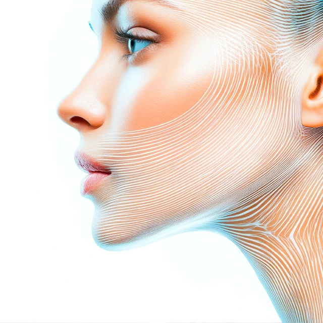
Jaw Asymmetry Surgery : The surgical approach to facial harmony
The human face is rarely perfectly symmetrical, and minor discrepancies are common and often go unnoticed. However, when these asymmetries become pronounced, particularly in the jaw structure, they can lead to significant functional impairments, aesthetic concerns, and psychological distress. Surgical correction of jaw asymmetry, a complex and highly specialized field within oral and maxillofacial surgery, aims to restore facial balance, optimize dental occlusion, and improve overall quality of life for affected individuals. This comprehensive overview will delve into the multifaceted aspects of jaw asymmetry, from its diverse etiologies and diagnostic methodologies to the intricate surgical planning, techniques, and post-operative care involved in its correction.
Jaw asymmetry refers to a noticeable lack of proportionality between the two sides of the lower face, specifically involving the mandible (lower jaw) and often extending to the maxilla (upper jaw), dental arches, and associated soft tissues. This imbalance can manifest in various ways, including:
- Mandibular deviation: The chin and lower jaw are shifted to one side.
- Occlusal cant: The biting plane of the teeth is tilted.
- Midline discrepancies: The dental midlines (between the central incisors) do not align with each other or the facial midline.
- Soft tissue asymmetry: Differences in muscle bulk, fat distribution, or skin folds on either side of the face.
- Condylar asymmetry: Discrepancies in the size, shape, or position of the temporomandibular joint (TMJ) condyles.
The scope of jaw asymmetry correction extends beyond mere aesthetics, encompassing the restoration of proper masticatory function, speech articulation, and TMJ health.
Prevalence and Impact
While mild facial asymmetry is universal, clinically significant jaw asymmetry is less common but affects a considerable number of individuals. Its prevalence varies depending on the definition and diagnostic criteria used, but it is a frequent concern in orthognathic surgery practices.
The impact of jaw asymmetry can be profound:
- Functional: Malocclusion can lead to inefficient chewing, difficulty biting, and uneven wear of teeth. TMJ dysfunction, including pain, clicking, and limited mouth opening, is a common co-morbidity. Speech impediments can also arise.
- Aesthetic: The visible imbalance can significantly affect a person’s self-image and confidence. Facial asymmetry is often perceived as less attractive, leading to social anxiety and reduced self-esteem.
- Psychological: Patients often report feelings of self-consciousness, embarrassment, and even depression due to their facial appearance. The psychological burden can be as significant as, if not more than, the functional limitations.
Etiology (Causes) of Jaw Asymmetry
Jaw asymmetry can originate from a variety of factors, broadly categorized as congenital, developmental, or acquired. Understanding the etiology is crucial for accurate diagnosis and effective treatment planning.
Congenital Causes
These are present at birth, often due to genetic factors or disturbances during embryonic development.
- Hemifacial Microsomia (HFM): A spectrum of deformities affecting the first and second branchial arches, resulting in underdevelopment of the ear, mandible, maxilla, zygoma, and associated soft tissues on one side of the face. The severity can range from mild ear anomalies to severe facial skeletal and soft tissue deficiencies.
- Goldenhar Syndrome (Oculo-auriculo-vertebral dysplasia): A more severe form of HFM, often including ocular (eye) and vertebral (spinal) anomalies.
- Treacher Collins Syndrome (Mandibulofacial Dysostosis): Characterized by underdeveloped facial bones, particularly the cheekbones and jaw, often with downward-slanting eyes, sparse eyelashes, and ear deformities. While often bilateral, it can present with significant asymmetry.
- Cleft Lip and Palate-related Deformities: Unilateral clefts can lead to maxillary hypoplasia and associated mandibular asymmetry due to altered growth patterns and scar tissue contracture.
Developmental Causes
These asymmetries develop during the growth period, often becoming apparent during adolescence.
- Condylar Hyperplasia (CH): An overgrowth of one mandibular condyle, leading to progressive elongation of the affected side of the mandible. This results in chin deviation to the contralateral (unaffected) side, an occlusal cant, and often a posterior open bite on the affected side. CH is classified into:
- Type 1 (Hemimandibular Hyperplasia): Characterized by increased condylar head and neck size, leading to elongation of the entire mandibular body on the affected side.
- Type 2 (Hemimandibular Elongation): Primarily involves elongation of the condylar neck and ramus, with less involvement of the condylar head or mandibular body.
- Condylar Hypoplasia: Underdevelopment of one mandibular condyle, often resulting from trauma or infection in childhood, leading to a shorter ramus and body on the affected side, with chin deviation towards the affected side.
- Idiopathic Condylar Resorption (ICR): A progressive and often bilateral condition, but can be asymmetric, leading to destruction and shortening of the condylar head. More common in young females, it results in an anterior open bite and progressive retrusion of the mandible. If unilateral, it causes severe asymmetry.
- Unilateral Macrognathia/Micrognathia: Generalized overgrowth or undergrowth of one side of the entire mandible, not necessarily originating from the condyle.
- Unilateral Maxillary Hypoplasia/Hyperplasia: Asymmetric growth of the upper jaw, leading to occlusal cant and midface asymmetry.
Acquired Causes
These occur after growth is complete or due to external factors.
- Trauma:
- Condylar Fractures: Untreated or poorly managed condylar fractures, especially in children, can disrupt growth or lead to fibrous ankylosis, resulting in mandibular asymmetry.
- Facial Bone Fractures: Fractures of the maxilla, zygoma, or orbital bones can lead to secondary soft tissue and skeletal asymmetry if not perfectly reduced.
- Temporomandibular Joint (TMJ) Ankylosis: Fusion of the condyle to the glenoid fossa, restricting jaw movement. If unilateral, it can severely impede growth on the affected side in children, leading to profound asymmetry. In adults, it causes functional limitation and can lead to secondary remodeling.
- Tumors: Benign or malignant tumors affecting the mandible, maxilla, or surrounding soft tissues can cause localized overgrowth, destruction, or displacement, leading to asymmetry.
- Infections: Chronic osteomyelitis or other severe infections can lead to bone destruction and asymmetry.
- Iatrogenic Causes: Asymmetry resulting from previous surgical interventions, such as poorly executed orthognathic surgery or tumor resections without adequate reconstruction.
Anatomy and Physiology Relevant to Jaw Asymmetry
A thorough understanding of the anatomical structures and their physiological functions is paramount for diagnosing and surgically correcting jaw asymmetry.
Skeletal Components
The primary skeletal components involved in jaw asymmetry are the maxilla, mandible, zygoma, and the cranial base.
- Maxilla: The upper jaw bone, forming the central part of the midface. Its position dictates the upper dental arch and contributes significantly to facial width and vertical dimension. Asymmetry here often manifests as an occlusal cant.
- Mandible: The lower jaw, the only movable bone of the face. Its condyles articulate with the temporal bone at the TMJs. Mandibular asymmetry is the most common form of jaw asymmetry, involving deviations of the chin, body, ramus, and condyle.
- Zygoma (Cheekbones): These bones contribute to facial width and projection. Asymmetry can be primary or secondary to mandibular/maxillary issues.
- Cranial Base: The base of the skull, which provides the foundation for the facial skeleton. Asymmetries in the cranial base can influence facial growth and contribute to overall facial imbalance.
Muscular Components
The muscles of mastication and facial expression play a crucial role in both the development and functional consequences of jaw asymmetry.
- Muscles of Mastication:
- Masseter, Temporalis, Medial Pterygoid: Primarily responsible for closing the jaw.
- Lateral Pterygoid: Primarily responsible for opening the jaw, protrusion, and lateral movements.
- Asymmetry can lead to hypertrophy (enlargement) of muscles on one side due to compensatory function or disuse atrophy on the other. This contributes to soft tissue asymmetry and can influence skeletal remodeling over time.
- Facial Mimetic Muscles: These muscles control facial expressions. Asymmetry can affect the symmetry of the mouth, eyes, and overall facial animation.
Temporomandibular Joint (TMJ)
The TMJ is a complex synovial joint connecting the mandible to the skull. It is critical for jaw movement and is frequently involved in jaw asymmetry.
- Structure: Composed of the mandibular condyle, the articular fossa of the temporal bone, and an articular disc separating these two components.
- Function: Allows for various movements, including rotation and translation, essential for chewing, speaking, and yawning.
- Role in Asymmetry:
- Condylar Pathology: Conditions like condylar hyperplasia, hypoplasia, or idiopathic condylar resorption directly cause or contribute to asymmetry.
- Adaptive Changes: The TMJ can undergo adaptive remodeling in response to long-standing asymmetry, which can further complicate treatment.
- Dysfunction: Asymmetry often leads to TMJ dysfunction, characterized by pain, clicking, popping, limited opening, or locking.
Dental Occlusion
Dental occlusion refers to the way the upper and lower teeth meet when the jaws are closed.
- Ideal Occlusion: A balanced relationship where teeth intercuspate efficiently, distributing forces evenly.
- Malocclusion in Asymmetry: Jaw asymmetry almost invariably results in malocclusion, such as:
- Crossbite: Upper teeth bite inside the lower teeth on one side.
- Open bite: A vertical gap between upper and lower teeth when the jaws are closed.
- Midline discrepancy: The dental midlines do not align.
- Occlusal cant: The plane of occlusion is tilted.
Correcting the occlusion is a primary goal of surgical intervention, as it is fundamental for functional rehabilitation.
Diagnosis and Assessment of Jaw Asymmetry
Accurate diagnosis and meticulous assessment are the cornerstones of successful surgical correction. This involves a systematic approach combining patient history, clinical examination, and advanced imaging.
Comprehensive Patient History
A detailed history helps identify the etiology, progression, and patient concerns.
- Onset and Progression: When did the asymmetry first become noticeable? Has it been progressive (suggesting active growth like condylar hyperplasia) or stable?
- Associated Symptoms: Pain (facial, TMJ, headache), clicking/popping/locking of the TMJ, limited mouth opening, difficulty chewing, speech problems, nasal obstruction.
- Previous Trauma/Surgeries: History of facial fractures, previous orthodontic treatment, or orthognathic surgery.
- Family History: Any family members with similar facial deformities.
- Psychological Impact: Assess the patient’s concerns regarding aesthetics and self-esteem.
Clinical Examination
A thorough clinical examination includes both extraoral and intraoral assessments.
Extraoral Assessment
This evaluates the overall facial balance and specific features.
- Facial Proportions: Assess vertical facial thirds and horizontal facial fifths.
- Facial Symmetry:
- Midline Deviation: Evaluate the deviation of the nasal tip, philtrum, upper dental midline, lower dental midline, and chin point relative to the true facial midline.
- Canting: Observe the cant (tilt) of the interpupillary line, occlusal plane, and commissures (corners of the mouth). An occlusal cant is a hallmark of jaw asymmetry.
- Soft Tissue Fullness/Deficiency: Note any differences in muscle bulk (masseter), fat pads, or skin folds, which can contribute to or mask skeletal asymmetry.
- Mandibular Deviation: Observe the chin position at rest, during opening, and during closing. Does the mandible deviate to one side during opening?
- TMJ Examination: Palpate the TMJs for tenderness, crepitus (grating sounds), or clicking during movement. Assess the range of motion (maximal incisal opening, lateral excursions).
- Neurological Assessment: Briefly check facial nerve (VII) function for any paresis or paralysis, especially if a congenital or acquired neurological condition is suspected.
Intraoral Assessment
This focuses on dental occlusion and oral health.
- Dental Occlusion:
- Angle’s Classification: Determine the Angle’s classification of malocclusion (Class I, II, or III) on both sides.
- Crossbite: Identify any anterior or posterior crossbites.
- Open Bite: Note any anterior or posterior open bites.
- Dental Midline Discrepancy: Measure the deviation of the upper and lower dental midlines.
- Occlusal Plane Cant: Visually assess the cant of the occlusal plane relative to the interpupillary line.
- Dental Health: Check for caries, periodontal disease, and existing restorations.
- Oral Mucosa and Tongue: Inspect for any abnormalities.
Imaging Studies
Advanced imaging is indispensable for visualizing the skeletal and soft tissue structures in three dimensions.
2D Radiography
- Panoramic Radiograph (OPG): Provides an overview of the entire mandible, maxilla, and dentition. Useful for assessing condylar morphology, ramus height, and dental status.
- Cephalometric Radiographs:
- Lateral Cephalogram: Assesses anteroposterior and vertical skeletal relationships. Limited for asymmetry as it’s a 2D projection of a 3D structure.
- Posterior-Anterior (PA) Cephalogram: Crucial for initial assessment of transverse asymmetry. Allows measurement of mandibular body length, ramus height, and condylar positions relative to the midline.
- TMJ Radiographs: Transcranial or transcervical views offer limited information but can show gross condylar pathology. Largely superseded by 3D imaging.
3D Imaging (Crucial for Asymmetry)
These modalities provide detailed, volumetric data, essential for accurate diagnosis and surgical planning.
- Cone Beam Computed Tomography (CBCT): The gold standard for assessing jaw asymmetry.
- Detailed Skeletal Assessment: Provides high-resolution images of the maxilla, mandible, zygoma, and cranial base, allowing precise measurement of bone dimensions, angles, and volumes.
- Condylar Morphology: Excellent for evaluating condylar size, shape, position, and any degenerative changes or hyperplasia.
- Volumetric Analysis: Allows for quantitative assessment of bone volume discrepancies.
- Airway Assessment: Can evaluate the pharyngeal airway space, which may be affected by jaw position.
- Computed Tomography (CT): Similar to CBCT but typically higher radiation dose and less ideal for dental structures. Used for very complex cases, tumor assessment, or when soft tissue detail is also critical.
- Magnetic Resonance Imaging (MRI): Primarily used for soft tissue assessment, particularly for the TMJ.
- TMJ Disc Position: Visualizes the articular disc, crucial for diagnosing disc displacement.
- Soft Tissue Pathology: Detects inflammation, effusions, or other soft tissue lesions within the TMJ.
Bone Scintigraphy (Technetium-99m)
- Purpose: Used to detect active bone growth, particularly in cases of suspected condylar hyperplasia. Increased uptake of the radioactive tracer in one condyle indicates active growth, guiding the timing of surgery (often waiting until growth ceases or performing a condylectomy if active).
Diagnostic Models and Articulation
- Dental Impressions and Stone Models: Provide a physical representation of the dental arches.
- Facebow Transfer and Articulator Mounting: Allows the dental models to be oriented in a mechanical articulator, simulating the patient’s jaw movements and occlusal relationships. This is critical for pre-surgical orthodontic planning and predicting occlusal changes.
- Virtual Surgical Planning (VSP): This advanced technology has revolutionized the treatment of jaw asymmetry and will be discussed in detail below.
Treatment Planning for Jaw Asymmetry
The correction of jaw asymmetry demands a highly individualized and multidisciplinary approach, integrating the expertise of several specialists to achieve optimal functional and aesthetic outcomes.
Multidisciplinary Approach
Successful treatment relies on close collaboration among:
- Orthodontist: Responsible for pre-surgical and post-surgical tooth alignment.
- Oral and Maxillofacial Surgeon (OMFS): Performs the surgical osteotomies and repositioning of the jaw bones.
- Prosthodontist: May be involved if extensive dental restorations or implants are needed.
- Ear, Nose, and Throat (ENT) Surgeon: If nasal or airway issues are present.
- Plastic Surgeon: For adjunctive soft tissue procedures (e.g., fat grafting, rhytidectomy).
- Psychologist/Counselor: To address the psychological impact of asymmetry and manage patient expectations.
Goals of Treatment
The primary goals are comprehensive:
- Functional Improvement:
- Establish a stable and functional dental occlusion.
- Improve masticatory efficiency and comfort.
- Enhance speech articulation.
- Optimize TMJ health and reduce symptoms.
- Improve airway patency if compromised.
- Aesthetic Balance:
- Achieve facial symmetry in all three dimensions (anteroposterior, vertical, transverse).
- Correct chin deviation and occlusal cant.
- Improve overall facial harmony and proportion.
- Psychological Well-being:
- Boost self-confidence and self-esteem.
- Alleviate social anxiety and body image concerns.
Phased Treatment Strategy
The correction of jaw asymmetry typically follows a well-defined sequence.
1. Pre-Surgical Orthodontics
This phase is crucial and can last from 12 to 24 months.
- Decompensation: The orthodontist aligns the teeth over their respective basal bones, often making the malocclusion appear worse initially. This is necessary to allow for maximal skeletal correction during surgery. For example, teeth that have compensated for a jaw deviation will be moved to their ideal positions on each jaw, creating a larger discrepancy between the upper and lower arches that can only be resolved surgically.
- Arch Coordination: Ensuring the upper and lower dental arches are properly shaped and sized to fit together after surgery.
- Creation of Stable Occlusal Platforms: Preparing the teeth to interdigitate perfectly into the surgical splint after surgery, providing a stable guide for jaw repositioning.
2. Surgical Phase
This is the core of the treatment, performed by the OMFS. It involves osteotomies (bone cuts) to reposition the maxilla, mandible, and chin.
- Maxillary Surgery (Lefort I Osteotomy): Used to correct maxillary cant, transverse discrepancies, and vertical excesses or deficiencies.
- Mandibular Surgery (Bilateral Sagittal Split Osteotomy – BSSO, Vertical Ramus Osteotomy – VRO, Inverted L/C Osteotomy): Used to reposition the mandible, correct chin deviation, and address condylar issues.
- Genioplasty: Chin surgery to further refine chin position, symmetry, and projection.
- Adjunctive Procedures: May include zygomatic osteotomies, orbital rim contouring, soft tissue grafting, or prosthetic implants to address residual skeletal or soft tissue asymmetries.
3. Post-Surgical Orthodontics
This phase typically lasts 6-12 months.
- Fine-tuning Occlusion: Minor adjustments to achieve the final, ideal bite.
- Retention: Use of retainers (fixed or removable) to maintain the corrected tooth and jaw positions long-term.
Virtual Surgical Planning (VSP)
Virtual Surgical Planning has revolutionized the precision and predictability of orthognathic surgery, particularly for complex asymmetries.
- Data Acquisition:
- CBCT Scan: Provides a 3D skeletal model of the patient’s skull.
- Intraoral Scan: Creates a digital 3D model of the patient’s teeth and dental arches.
- These two datasets are then fused digitally.
- 3D Reconstruction and Segmentation: Specialized software reconstructs the patient’s anatomy in 3D. Individual bones (maxilla, mandible, zygoma, etc.) are segmented (separated) virtually.
- Surgical Simulation: The surgeon, often in collaboration with the orthodontist and VSP engineers, performs virtual osteotomies and repositions the bone segments in a 3D environment.
- This allows for precise measurement of bone movements in all three planes.
- The surgeon can visualize the exact skeletal changes and predict the resulting occlusal relationship.
- Multiple surgical plans can be tested and refined.
- Prediction of Soft Tissue Changes: Some advanced VSP software can provide a reasonable prediction of how the soft tissues will drape over the new skeletal framework.
- Design and Fabrication of Surgical Splints and Guides:
- Intermediate Splint: Designed to guide the repositioning of the maxilla relative to the cranial base, or the mandible relative to the maxilla, during surgery.
- Final Splint: Guides the final interdigitation of the upper and lower teeth.
- Custom Cutting Guides: For complex osteotomies (e.g., segmental osteotomies, precise genioplasty cuts), custom guides can be 3D printed to ensure exact bone cuts.
- Custom Plates: In some highly asymmetric cases, custom-bent or custom-designed fixation plates can be fabricated pre-operatively based on the VSP.
- Benefits of VSP:
- Enhanced Precision and Predictability: Minimizes intraoperative guesswork and improves the accuracy of bone repositioning.
- Reduced Operative Time: Pre-planning streamlines the surgical procedure.
- Improved Communication: Facilitates clear communication among the surgical team and with the patient.
- Optimized Outcomes: Leads to more stable, functional, and aesthetically pleasing results.
- Patient Education: Patients can visualize their planned surgical outcome, improving understanding and managing expectations.
Surgical Techniques for Correction of Jaw Asymmetry
The surgical phase involves a combination of osteotomies, carefully chosen and executed to address the specific skeletal discrepancies identified during planning. All intraoral osteotomies are performed through incisions made inside the mouth, leaving no visible external scars.
Maxillary Osteotomies (Lefort I Osteotomy)
- Indications for Asymmetry:
- Maxillary Cant: To level the occlusal plane of the upper jaw.
- Transverse Deficiency/Excess: To widen or narrow the upper dental arch.
- Vertical Excess/Deficiency: To impact (shorten) or down-graft (lengthen) the maxilla.
- Technique:
- An incision is made in the gum above the upper teeth.
- The maxilla is exposed, and horizontal osteotomy cuts are made above the tooth roots, extending from the piriform aperture (nasal opening) posteriorly to the pterygomaxillary junction.
- The maxilla is then down-fractured (separated from the skull base).
- Specific Considerations for Asymmetry:
- Differential Impaction/Down-grafting: To correct an occlusal cant, more bone is removed from the side that needs to be impacted, or bone grafts are placed on the side that needs to be lengthened.
- Segmental Osteotomies: In some cases, the maxilla might be cut into multiple segments to correct severe transverse discrepancies or localized canting.
- The maxilla is repositioned into its planned new position using the intermediate surgical splint as a guide.
- Rigid fixation is achieved using titanium plates and screws.
Mandibular Osteotomies
These procedures are used to reposition the lower jaw.
1. Bilateral Sagittal Split Osteotomy (BSSO)
- Indications for Asymmetry: The most versatile and commonly performed mandibular osteotomy for correcting a wide range of mandibular asymmetries, including advancement, setback, rotation, and correction of chin deviation.
- Technique:
- Incisions are made inside the mouth along the anterior border of the mandibular ramus.
- Osteotomy cuts are made through the outer (buccal) cortical bone, extending along the lateral ramus and then horizontally along the medial aspect of the ramus, behind the lingula (where the inferior alveolar nerve enters).
- The inner (medial) cortical plate is then split from the outer (lateral) cortical plate, effectively « splitting » the ramus into two segments.
- The distal (tooth-bearing) segment of the mandible is then moved to its planned position.
- Specific Considerations for Asymmetry:
- Differential Movements: One side of the mandible may be advanced or set back more than the other, or rotated differently, to achieve symmetry.
- Condylar Seating: Meticulous attention is paid to ensuring proper seating of the condyles in the glenoid fossae to prevent post-operative TMJ issues and relapse.
- Rigid fixation is achieved with titanium plates and screws.
2. Vertical Ramus Osteotomy (VRO)
- Indications: Primarily used for mandibular setback, especially in cases where there’s a need to avoid the inferior alveolar nerve, or in cases of severe condylar hyperplasia where a condylectomy is also performed. Less commonly used for complex 3D asymmetry correction due to less precise control.
- Technique: A vertical cut is made through the mandibular ramus, typically from the sigmoid notch to the angle of the mandible. The proximal (condylar) segment is allowed to overlap the distal segment. Fixation is usually achieved through muscle and soft tissue tension, or sometimes with wires.
3. Inverted L/C Osteotomy
- Indications: Used for significant mandibular length discrepancies, particularly in cases of severe condylar hyperplasia or hypoplasia, or for reconstruction after trauma/tumor resection. Allows for significant lengthening or shortening of one side of the mandible.
- Technique: An L-shaped or C-shaped osteotomy is performed in the ramus and/or body of the mandible. Bone grafts can be interposed to lengthen the mandible, or bone can be removed to shorten it. Fixation is with plates and screws.
Condylar Management
- Condylectomy/Condylotomy: For active condylar hyperplasia, a high condylectomy (removal of the actively growing part of the condyle) or a condylar shave (contouring the condyle) may be performed to stop the overgrowth. This is often done prior to or concurrently with orthognathic surgery.
- TMJ Reconstruction: In cases of severe degenerative changes, ankylosis, or condylar defects (e.g., after tumor resection), the TMJ may need to be reconstructed using autogenous grafts (e.g., costochondral graft from the rib) or alloplastic (artificial) implants.
Genioplasty (Chin Surgery)
- Indications for Asymmetry: Almost always performed in conjunction with orthognathic surgery for jaw asymmetry to achieve final chin symmetry, projection, and vertical height. The chin is often deviated to the same side as the mandibular body deviation.
- Technique:
- An incision is made inside the lower lip.
- A horizontal osteotomy cut is made through the lower border of the mandible, below the tooth roots.
- The mobilized chin segment is then repositioned in three dimensions:
- Transverse: Moved laterally to correct deviation.
- Anteroposterior: Advanced or set back for projection.
- Vertical: Shortened or lengthened.
- Rotation: Rotated to correct cant.
- The chin segment is fixed with titanium plates and screws.
Adjunctive Procedures
To optimize facial symmetry, other procedures may be performed.
- Zygomatic Osteotomies/Contouring: To address asymmetry in the cheekbones.
- Orbital Rim Contouring: To correct orbital asymmetry.
- Soft Tissue Augmentation: Fat grafting or dermal fillers can be used to camouflage minor skeletal deficiencies or augment soft tissue volume on the deficient side.
- Soft Tissue Reduction: Buccal fat pad removal or masseter reduction (e.g., with Botox) can be used on the hypertrophic side.
- Prosthetic Implants: Custom-designed implants (e.g., for mandibular angle or malar region) can be used to augment areas of skeletal deficiency.
Post-Operative Management and Recovery
The post-operative period is critical for healing, stability, and achieving the final outcome.
Immediate Post-Op
- Swelling: Significant facial swelling is expected and peaks around 48-72 hours. Ice packs, head elevation, and sometimes corticosteroids are used to manage this.
- Pain: Managed with prescribed analgesics.
- Nausea: Common due to anesthesia and medications, managed with antiemetics.
- Bleeding: Minor oozing is normal; significant bleeding is rare.
- Airway Management: Close monitoring, especially for mandibular advancement surgeries, as swelling can temporarily compromise the airway. Rarely, an overnight stay in an intensive care unit might be necessary.
Diet Progression
- Liquid Diet: For the first 1-2 weeks to allow initial bone healing.
- Soft Non-Chewing Diet: For the next 4-6 weeks. Patients are instructed not to chew anything hard.
- Gradual Return to Normal Diet: Over several months, as bone healing progresses.
Oral Hygiene
- Gentle Brushing: With a soft toothbrush.
- Antiseptic Rinses: Chlorhexidine mouthwash is often prescribed to reduce bacterial load and promote healing.
Elastics and Fixation
- Intermaxillary Fixation (IMF): While rigid internal fixation (plates and screws) has largely replaced prolonged IMF, some surgeons may use light elastics or temporary wiring for a short period (days to weeks) to guide the bite and stabilize the segments.
- Orthodontic Elastics: Used post-operatively to guide the occlusion into its final position and to encourage muscle adaptation.
Physical Therapy
- Jaw Exercises: Gentle exercises to restore range of motion are initiated once approved by the surgeon, typically a few weeks post-op.
- TMJ Rehabilitation: If TMJ dysfunction was present pre-operatively or develops post-operatively.
Follow-up Schedule
Regular follow-up appointments with both the surgeon and orthodontist are essential to monitor healing, assess occlusion, and manage any complications.
Potential Complications
While orthognathic surgery is generally safe, potential complications exist.
- General Surgical Risks:
- Bleeding: Intraoperative or post-operative hemorrhage.
- Infection: At the surgical sites or around the fixation hardware.
- Anesthesia Risks: Allergic reactions, respiratory complications.
- Adverse Drug Reactions.
- Specific Orthognathic Risks:
- Nerve Injury:
- Inferior Alveolar Nerve (IAN): Temporary or, rarely, permanent numbness (paresthesia/dysesthesia) of the lower lip, chin, and teeth on the affected side after BSSO.
- Mental Nerve: Sensory nerve to the lower lip and chin, can be affected during genioplasty.
- Facial Nerve (VII): Extremely rare, but can cause temporary or permanent weakness of facial muscles.
- Relapse: Skeletal or occlusal instability, where the jaws drift back towards their original position. Minimized by rigid fixation and proper planning.
- Non-union/Malunion: Failure of the bone segments to heal or healing in an incorrect position. Rare with modern techniques.
- TMJ Dysfunction: New onset or worsening of TMJ pain, clicking, or limited opening.
- Airway Compromise: Especially with mandibular setback, though usually transient.
- Aesthetic Dissatisfaction: Despite meticulous planning, patients may have unrealistic expectations or subtle residual asymmetry.
- Condylar Resorption: Post-surgical resorption of the condyles, particularly in patients predisposed to idiopathic condylar resorption.
- Hardware-related Issues: Infection, loosening, or palpability of plates and screws, sometimes requiring removal.
- Nerve Injury:
Long-Term Outcomes and Prognosis
The long-term success of jaw asymmetry correction is measured by the stability of the skeletal and occlusal results, functional improvements, aesthetic enhancements, and overall patient satisfaction.
Stability of Results
- Factors Influencing Stability:
- Rigid Fixation: Titanium plates and screws provide immediate stability, reducing relapse.
- Muscle Adaptation: Muscles of mastication adapt to the new jaw position.
- Condylar Health: Healthy TMJs are crucial for long-term stability. Pre-existing TMJ pathology or post-surgical condylar resorption can compromise results.
- Accurate Planning and Execution: Meticulous VSP and precise surgical execution are paramount.
- Most orthognathic surgery results are highly stable over many years, especially with modern techniques.
Functional Improvements
- Mastication: Patients typically experience a significant improvement in chewing efficiency and comfort.
- Speech: Articulation often improves as the dental and jaw relationships are normalized.
- Breathing: Airway patency can improve, particularly in cases of mandibular advancement, potentially reducing snoring or sleep apnea.
- TMJ Function: Many patients experience resolution or significant reduction of pre-existing TMJ symptoms.
Aesthetic Improvements
- Facial Symmetry: The most noticeable and desired outcome is a significant improvement in facial balance and symmetry.
- Profile Balance: Correction of chin deviation and establishment of harmonious facial proportions.
- Self-Esteem: The positive aesthetic changes often lead to a profound improvement in the patient’s self-confidence and psychological well-being.
Patient Satisfaction
Studies consistently show high levels of patient satisfaction following surgical correction of jaw asymmetry, reflecting the significant functional and aesthetic benefits achieved.
Need for Revision Surgery
While most cases achieve excellent results, a small percentage of patients may require revision surgery for residual asymmetry, relapse, or complications. This is typically due to complex initial deformities, unexpected healing, or rare complications.