

Ventre relâché ? Découvrez l’effet tenseur de l’Endolift
9 janvier 2026
Cinderella Feet Surgery : The Skinny on Skinny Feet
14 janvier 2026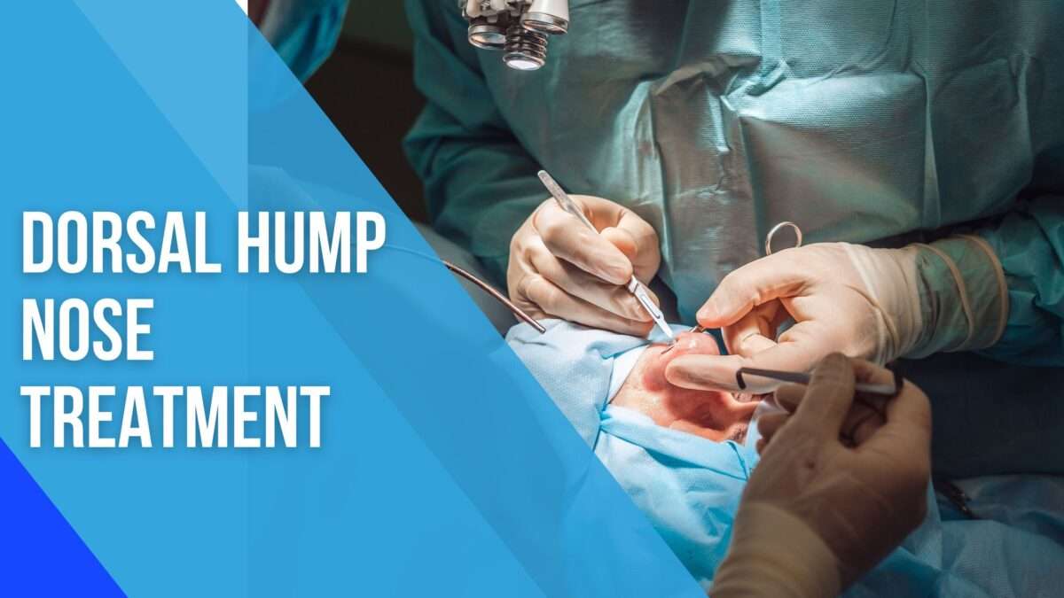
Nasal Hump Removal Surgery : Enhancing Facial Harmony
A « nasal hump » refers to a convexity or prominence along the bridge of the nose, often perceived as a bump or dorsal convexity. This feature can be composed of bone, cartilage, or a combination of both. While a prominent nasal dorsum is a natural variation of human anatomy and is often a distinguishing ethnic trait, for many individuals, it can be a source of self-consciousness, impacting facial harmony and overall self-perception.
Nasal hump removal surgery is a core component of rhinoplasty, the surgical reshaping of the nose. The primary objective is to reduce the height of the dorsal hump, creating a straighter, more refined nasal profile that better complements the patient’s other facial features. Beyond mere reduction, the procedure often involves a series of complementary maneuvers to ensure a natural-looking result, maintain nasal function, and prevent undesirable post-operative deformities. This is not just about « shaving down a bump »; it’s about sculpting the nose in three dimensions to achieve aesthetic balance while preserving or improving nasal breathing.
Anatomy of the Nose Relevant to Hump Removal
A thorough understanding of nasal anatomy is paramount for successful and safe dorsal hump reduction. The nasal dorsum, or bridge, is formed by a complex interplay of skeletal and soft tissue structures.
- Nasal Bones: The upper third to half of the nasal dorsum is composed of the paired nasal bones. These flat, rectangular bones articulate superiorly with the frontal bone and laterally with the maxilla. The junction of the nasal bones with the frontal bone forms the nasion, the deepest point of the nasal root.
- Upper Lateral Cartilages (ULCs): Inferior to the nasal bones, the dorsal hump is primarily cartilaginous, formed by the paired upper lateral cartilages. These triangular-shaped cartilages are intimately fused with the dorsal septum in the midline and articulate laterally with the nasal bones and the pyriform aperture. The caudal (lower) edges of the ULCs form the internal nasal valve, a critical area for airflow.
- Septal Cartilage (Dorsal Septum): The quadrangular cartilage of the nasal septum forms the central supporting structure of the nose. Its dorsal edge contributes significantly to the height and shape of the cartilaginous dorsum. A prominent dorsal septum is often a major component of the cartilaginous hump.
- Skin and Soft Tissue Envelope (S-STE): Overlying these skeletal and cartilaginous structures is the skin and soft tissue envelope, which includes skin, subcutaneous fat, and muscles (e.g., procerus, nasalis). The thickness and quality of the S-STE significantly influence the final aesthetic outcome, as a thin S-STE will reveal underlying irregularities more readily than a thick one.
- Nasal Airway: The internal nasal valve, formed by the caudal edge of the ULCs, the dorsal septum, and the head of the inferior turbinate, is the narrowest point of the nasal airway. Any surgical maneuver that compromises this area can lead to post-operative breathing difficulties.
The hump itself is typically a combination of excess bone from the nasal bones and excess cartilage from the ULCs and the dorsal septum. The proportion of bone to cartilage in the hump varies among individuals.
Indications for Nasal Hump Removal
The primary indication for nasal hump removal is aesthetic dissatisfaction with a prominent nasal dorsum. Patients often describe their nose as « too big, » « hooked, » or having a « bump. »
- Aesthetic Concerns:
- Dorsal Convexity: The most common reason, where the nasal bridge protrudes excessively, disrupting the smooth line from the glabella (area between eyebrows) to the nasal tip.
- Lack of Facial Harmony: A prominent hump can overpower other facial features, making the nose appear disproportionately large or out of balance.
- Profile Imbalance: When viewed in profile, a hump can create an undesirable shadow or break in the aesthetic line.
- Gender-Specific Aesthetics: While a subtle dorsal convexity can be considered attractive in some male profiles, a prominent hump is generally less desired in female profiles, where a straighter or slightly concave dorsum is often preferred.
- Psychological Impact: For many, the aesthetic concern translates into significant psychological distress, affecting self-esteem, social interactions, and body image.
- Functional Considerations: While less common for an isolated hump, in some cases, a severe dorsal deviation or a very high hump can contribute to internal nasal valve collapse or airflow obstruction, though this is usually secondary to septal deviation. When functional issues coexist, they are addressed concurrently.
- Patient Expectations: A crucial indication is the patient’s clear, realistic understanding of the surgical goals and potential outcomes, and their desire for the aesthetic change.
Contraindications and Cautions
Not every individual seeking nasal hump removal is a suitable candidate. Careful screening is essential to ensure patient safety and satisfaction.
- Unrealistic Expectations: Patients who expect perfection, believe surgery will solve all their life problems, or have an idealized, unattainable image of their nose are generally poor candidates.
- Body Dysmorphic Disorder (BDD): Individuals with BDD have a preoccupation with perceived flaws in their appearance, which are often minimal or imagined by others. Surgery rarely satisfies these patients and can exacerbate their distress. A psychological evaluation is crucial if BDD is suspected.
- Active Infections: Any active infection, particularly in the nasal or facial area, must be resolved before surgery.
- Certain Medical Conditions:
- Uncontrolled Chronic Diseases: Uncontrolled diabetes, severe hypertension, or significant cardiovascular disease increase surgical risks.
- Bleeding Disorders: Hemophilia or other coagulopathies are significant contraindications due to the risk of excessive bleeding. Patients on anticoagulants must discontinue them pre-operatively under medical supervision.
- Autoimmune Diseases: While not an absolute contraindication, these require careful consideration due to potential impacts on healing and immune response.
- Age Considerations:
- Skeletal Immaturity: Rhinoplasty is generally postponed until nasal skeletal growth is complete, typically around 15-16 for girls and 16-17 for boys. Operating on an immature nose can disrupt growth and lead to unpredictable results.
- Smoking: Smoking significantly impairs wound healing, increases the risk of infection, and can compromise blood supply to tissues. Patients are strongly advised to cease smoking several weeks before and after surgery.
- Previous Nasal Surgery (Revision Cases): While not a contraindication, revision rhinoplasty is significantly more complex due to altered anatomy, scar tissue, and often diminished cartilage resources. It requires a highly experienced surgeon.
- Psychological Instability: Patients undergoing significant life stress, depression, or other mental health crises may not be in the best state to make informed decisions about elective cosmetic surgery.
Pre-operative Assessment and Planning
The success of nasal hump removal hinges on meticulous pre-operative assessment and planning. This phase involves a comprehensive evaluation of the patient’s medical history, physical nasal characteristics, aesthetic goals, and psychological readiness.
Patient Consultation
The initial consultation is a critical dialogue between the patient and the surgeon.
- Detailed Medical History:
- Past Surgeries: Any previous nasal surgeries, facial trauma, or other relevant surgical history.
- Medical Conditions: Chronic illnesses, allergies (especially to medications or anesthesia), bleeding tendencies.
- Medications: Current prescriptions, over-the-counter drugs, herbal supplements (many can increase bleeding risk, e.g., aspirin, NSAIDs, Vitamin E, ginkgo biloba).
- Social History: Smoking, alcohol consumption, recreational drug use.
- Functional Concerns: History of nasal obstruction, difficulty breathing, chronic sinusitis, or epistaxis (nosebleeds).
- Psychological Assessment:
- Motivation: Understanding why the patient desires surgery. Is it for themselves or to please others?
- Expectations: Are their expectations realistic and aligned with what surgery can achieve?
- Emotional Stability: Screening for signs of BDD or other psychological issues.
- Discussion of Goals and Expectations: Open communication about what the patient likes and dislikes about their nose, and what they hope to achieve. The surgeon must clearly explain what is surgically possible and what limitations exist.
Physical Examination
A thorough physical examination of the nose and surrounding facial structures is essential.
- External Nasal Analysis:
- Skin Quality: Thickness, elasticity, presence of acne or telangiectasias.
- Facial Symmetry: Overall facial balance and proportion.
- Nasal Projection: How far the nose protrudes from the face.
- Nasal Rotation: The angle of the nasal tip relative to the upper lip.
- Dorsal Line: Assessment of the hump’s extent (bony vs. cartilaginous), its height, and its relationship to the nasion and tip.
- Tip Definition: Shape, width, and projection of the nasal tip.
- Alar Base Width: Width of the nostrils.
- Palpation: Assessment of the underlying bony and cartilaginous framework for stability, strength, and any deviations.
- Internal Nasal Examination:
- Speculum Examination: Evaluation of the nasal septum for deviation, spurs, or perforations.
- Turbinates: Assessment of their size and potential for hypertrophy, which can cause obstruction.
- Nasal Valves: Evaluation of both internal and external nasal valves for patency and potential collapse. This is crucial as dorsal hump reduction can sometimes compromise the internal nasal valve.
Photography
Standardized pre-operative photographs are indispensable. They serve multiple purposes:
- Baseline Documentation: Records the initial state of the nose.
- Surgical Planning: Allows for detailed analysis of nasal aesthetics from various angles (frontal, lateral, oblique, basal, worm’s eye).
- Communication Tool: Facilitates discussion with the patient about specific features.
- Post-operative Comparison: Essential for evaluating surgical outcomes.
Imaging (If Necessary)
While not routinely required for purely aesthetic hump reduction, certain situations may warrant imaging:
- CT Scan: For patients with a history of significant nasal trauma, complex septal deviations, chronic sinusitis, or when functional concerns are prominent. It provides detailed information about bony and cartilaginous anatomy.
Computer Imaging/Morphing
Many surgeons utilize computer imaging software during the consultation. This tool allows the surgeon to digitally manipulate the patient’s photographs to simulate potential post-operative outcomes.
- Benefits: Helps align patient and surgeon expectations, visualizes the impact of different degrees of hump reduction, and facilitates a shared understanding of the aesthetic goals.
- Caveat: It’s crucial to emphasize that computer imaging is a simulation and not a guarantee of the exact surgical outcome.
Informed Consent
A comprehensive informed consent process is mandatory. The surgeon must thoroughly discuss:
- The Procedure: Detailed explanation of what the surgery entails.
- Expected Outcomes: Realistic discussion of aesthetic improvements.
- Potential Risks and Complications: A detailed review of all possible adverse events, both general and specific to rhinoplasty.
- Alternatives to Surgery: Although limited for significant hump reduction, non-surgical options (e.g., fillers for camouflage) should be mentioned if applicable.
- Recovery Process: What to expect during the healing period.
- Cost: Full breakdown of all associated fees.
Surgical Techniques for Hump Removal
Nasal hump reduction is a delicate balance of subtraction and reconstruction. The goal is not just to remove the hump but to create a smooth, natural dorsal line while maintaining structural integrity and nasal function.
Surgical Approaches
There are two main surgical approaches to rhinoplasty, both of which can be used for hump reduction:
- Open Rhinoplasty:
- Incision: Involves a small, inverted V or W-shaped incision across the columella (the strip of skin between the nostrils), connecting to incisions inside the nostrils.
- Advantages: Provides direct, unobstructed visualization of the underlying bony and cartilaginous framework. This allows for precise and controlled removal of the hump and subsequent reconstruction. It is often preferred for complex cases or when significant structural changes are needed.
- Disadvantages: Leaves a small external scar (usually inconspicuous), and may result in slightly more initial swelling and a longer period of numbness in the nasal tip compared to closed rhinoplasty.
- Closed Rhinoplasty (Endonasal Rhinoplasty):
- Incision: All incisions are made inside the nostrils, meaning there is no external scar.
- Advantages: No external scar, potentially less initial swelling, and quicker recovery of sensation.
- Disadvantages: Limited visualization of the underlying structures, requiring the surgeon to work through small openings. This « tunnel vision » can make precise and symmetrical hump reduction more challenging, especially for larger humps or complex deformities. It relies heavily on the surgeon’s tactile feel and experience.
The choice of approach depends on the surgeon’s preference, training, and the complexity of the individual case. For significant hump reduction, open rhinoplasty often offers superior control and precision.
Hump Reduction Methods
Once the nasal framework is exposed (via open approach) or accessed (via closed approach), the hump can be reduced using various instruments:
- Osteotomes (Chisels): These surgical instruments are used to remove the bony component of the hump. They are typically sharp and come in various widths. The surgeon carefully positions the osteotome at the desired level of reduction and uses a mallet to gently tap it, precisely separating the excess bone. This is a controlled method for removing larger bony humps.
- Rasps: After the initial bony reduction with an osteotome, rasps (bone files) are used to meticulously smooth any remaining bony irregularities and achieve the desired dorsal contour. They are less aggressive than osteotomes and allow for fine-tuning.
- Surgical Saws: While less commonly used for routine hump reduction, specialized micro-oscillating saws can be employed for precise bone removal, particularly in revision cases or when dealing with very dense bone.
- Cartilage Scissors/Scalpel: The cartilaginous component of the hump (from the upper lateral cartilages and dorsal septum) is typically removed with a scalpel (e.g., #11 blade) or specialized cartilage scissors. This requires extreme precision to avoid over-resection and to maintain the integrity of the internal nasal valve.
- Powered Instruments: Some surgeons utilize powered instruments like micro-drills or burrs for very precise and controlled bone contouring, especially in revision cases or for smoothing.
The amount of hump removed is critical. Over-resection can lead to a « saddle nose deformity » (a scooped-out appearance) or an overly feminized nose in males. Under-resection leaves a residual hump, requiring revision. The goal is a natural, straight, or slightly curved dorsal line that harmonizes with the rest of the face.
Management of the Open Roof Deformity
A crucial step following hump removal is addressing the « open roof deformity. » When the bony and cartilaginous hump is removed, the nasal bones and upper lateral cartilages are separated, creating an open space on the dorsum. If left unaddressed, this can lead to:
- Widening of the Nasal Bridge: The nose appears broad and flat from the front.
- Irregularities: Visible gaps or steps along the dorsal line.
- Compromised Internal Nasal Valve: The upper lateral cartilages, now detached from the septum, can collapse inward, causing breathing difficulties.
To correct the open roof deformity, several techniques are employed:
- Osteotomies: These are controlled surgical fractures of the nasal bones, performed to narrow the nasal base and close the open roof.
- Lateral Osteotomies: Incisions are made inside the nostrils, and a narrow osteotome or saw is used to carefully fracture the nasal bones along their junction with the maxilla. This allows the surgeon to move the nasal bones medially (inward).
- Medial Osteotomies: Less common, these involve fracturing the nasal bones along their midline junction.
- Intermediate/Transverse Osteotomies: Sometimes used to mobilize the nasal bones more effectively.
Osteotomies are performed with extreme care to avoid damage to surrounding structures and to achieve symmetrical narrowing.
- Spreader Grafts: These are small strips of cartilage (typically harvested from the patient’s own septum, ear, or rib) that are placed between the dorsal septum and the upper lateral cartilages.
- Purpose:
- Maintain Internal Nasal Valve Patency: They push the upper lateral cartilages laterally, preventing their collapse and ensuring an adequate airway.
- Recreate Dorsal Aesthetic Lines: They help establish smooth, parallel lines along the nasal dorsum, preventing a « pinched » or « inverted V » deformity (where the lower edges of the nasal bones become visible).
- Camouflage Irregularities: They can help smooth out minor dorsal irregularities.
- Purpose:
- Camouflage Grafts: Tiny pieces of crushed cartilage or fascia can be used to fill in subtle depressions or smooth out minor irregularities along the dorsum, especially in patients with thin skin.
Associated Procedures
Dorsal hump reduction is rarely performed in isolation. To achieve a balanced and harmonious result, other rhinoplasty maneuvers are often performed concurrently:
- Tip Refinement: Often, reducing the dorsal hump makes the nasal tip appear relatively larger or less defined. Techniques like tip sutures (to reshape the tip cartilages) or tip grafts (small pieces of cartilage to add projection or definition) are commonly used.
- Septoplasty: If a deviated nasal septum contributes to breathing difficulties or asymmetry, it is corrected during the same procedure.
- Turbinate Reduction: Enlarged turbinates (structures inside the nose that warm and humidify air) can cause nasal obstruction. They can be reduced if necessary.
- Alar Base Reduction: If the nostrils appear too wide after dorsal reduction and tip refinement, a small wedge of tissue can be removed from the base of the nostrils to narrow them.
Anesthesia
Nasal hump removal surgery is typically performed under:
- General Anesthesia: The most common choice, where the patient is completely asleep and pain-free throughout the procedure. An anesthesiologist monitors vital signs continuously.
- Local Anesthesia with Intravenous (IV) Sedation: The patient is sedated but remains conscious, though deeply relaxed and unaware of the procedure. The nose is numbed with local anesthetic injections. This option may be suitable for less complex cases and offers a quicker recovery from anesthesia.
The choice of anesthesia is discussed with the patient and anesthesiologist, considering the patient’s health, preferences, and the complexity of the surgery.
The Surgical Procedure (Step-by-Step Overview)
While variations exist, a typical nasal hump removal procedure (using an open approach) follows these general steps:
- Anesthesia Administration: General anesthesia is induced.
- Infiltrative Anesthesia: A local anesthetic with a vasoconstrictor (e.g., lidocaine with epinephrine) is injected into the nasal tissues. This provides additional pain control, reduces bleeding, and aids in dissection.
- Incision and Dissection: For open rhinoplasty, the columellar incision is made, and the skin and soft tissue envelope is carefully lifted off the underlying bony and cartilaginous framework, exposing the entire dorsum, septum, and tip cartilages.
- Hump Marking: The surgeon carefully marks the precise amount of bone and cartilage to be removed, often using a marking pen and calipers to ensure accuracy and symmetry.
- Hump Removal:
- Cartilaginous Hump Reduction: The excess dorsal septal cartilage and upper lateral cartilages are carefully resected using a scalpel or scissors. This is often done first to visualize the bony hump more clearly.
- Bony Hump Reduction: An osteotome is precisely positioned at the base of the bony hump and gently tapped with a mallet to remove the desired amount of bone. Rasps are then used to smooth any remaining bony irregularities.
- Inspection and Refinement: The surgeon carefully inspects the newly contoured dorsum, ensuring a smooth, straight line. Any minor irregularities are addressed with further rasping or camouflage grafts.
- Management of the Open Roof:
- Osteotomies: Lateral osteotomies are performed to bring the nasal bones inward, closing the open roof deformity and narrowing the nasal bridge.
- Spreader Grafts: If indicated, spreader grafts are meticulously placed between the dorsal septum and the upper lateral cartilages and secured with sutures to maintain airway patency and dorsal aesthetic lines.
- Tip Refinement (if necessary): Sutures are placed in the tip cartilages, or cartilage grafts are positioned, to refine the nasal tip’s shape, projection, and rotation, ensuring it harmonizes with the newly reduced dorsum.
- Closure of Incisions: The columellar incision (in open rhinoplasty) and internal incisions are meticulously closed with fine sutures.
- Splinting:
- Internal Splints: Soft silicone splints may be placed inside the nostrils to support the septum and internal nasal valves, especially if septoplasty was performed.
- External Splint/Cast: A thermoplastic or plaster cast is applied to the outside of the nose. This provides external support, protects the reshaped bones and cartilages, and helps reduce swelling. Taping may also be used.
- Dressing: A sterile dressing is applied.
The entire procedure typically takes 2 to 4 hours, depending on the complexity and the number of associated procedures performed.
Post-operative Care and Recovery
The recovery period after nasal hump removal is crucial for optimal healing and results. Patients should be prepared for a gradual process.
Immediate Post-operative Period (First 24-48 Hours)
- Pain: Managed with prescribed oral pain medication. Pain is usually moderate and well-controlled.
- Swelling: Significant swelling of the nose and surrounding facial areas (cheeks, eyelids) is expected.
- Bruising: Black and blue bruising around the eyes is common and can extend to the cheeks.
- Nasal Packing: If internal packing was used (less common now with internal splints), it will be removed within 1-3 days.
- Drainage: Light bloody or serosanguinous drainage from the nostrils is normal.
- Head Elevation: Keeping the head elevated, even while sleeping, helps reduce swelling.
- Cold Compresses: Applied to the eyes and cheeks (not directly on the nasal cast) to minimize swelling and bruising.
- Activity Restriction: Strict rest is advised.
First Week
- Splint Removal: The external cast and any internal splints are typically removed by the surgeon around 5-7 days post-surgery.
- Initial Appearance: The nose will still be significantly swollen, and the skin may appear discolored. The initial shape will be visible, but it will not be the final result.
- Bruising Resolution: Bruising usually starts to fade, changing from purple to yellow/green.
- Sutures: Any external sutures (columellar) are removed. Internal sutures dissolve on their own.
- Nasal Congestion: Feeling of congestion is common due to internal swelling. Saline nasal sprays may be recommended.
- Activity: Light, non-strenuous activities can be resumed. Avoid bending, lifting, or anything that increases blood pressure.
Weeks 2-6
- Significant Swelling Reduction: Most of the noticeable swelling (especially around the eyes and upper nose) will subside. However, subtle swelling, particularly in the nasal tip, will persist for months.
- Return to Activities: Most normal daily activities, including work or school, can be resumed. Strenuous exercise, contact sports, and activities that could impact the nose must still be avoided.
- Glasses: Avoid wearing heavy glasses that rest on the nasal bridge for at least 4-6 weeks to prevent indentations or shifting of the bones. Lighter glasses or taping them to the forehead may be an option.
- Sun Protection: Protect the healing skin from direct sun exposure to prevent hyperpigmentation.
Months 3-12 (and Beyond)
- Gradual Resolution of Swelling: The nose will continue to refine as swelling slowly resolves. The tip is usually the last area to fully de-swell.
- Final Results Emerge: The true final results of the surgery typically become apparent around 12-18 months post-operatively, as all swelling has dissipated and tissues have settled.
- Numbness: Some degree of numbness in the nasal tip or dorsum is common and usually resolves over several months, though partial numbness can occasionally be permanent.
- Scar Maturation: Any external scar (columellar) will continue to fade and mature, becoming less noticeable over time.
General Post-operative Instructions
- Medications: Take prescribed antibiotics and pain relievers as directed.
- Nasal Hygiene: Gently clean the nostrils as instructed by the surgeon. Avoid aggressive blowing of the nose for several weeks.
- Diet: A soft, bland diet may be preferred initially.
- Sleeping: Continue to sleep with your head elevated.
- Avoid Trauma: Protect the nose from any bumps or impacts.
- Follow-up Appointments: Attend all scheduled post-operative appointments for monitoring and assessment.
- Psychological Adjustment: It’s normal to experience a range of emotions during recovery. Patience is key, as the final result takes time to manifest.
Potential Risks and Complications
While nasal hump removal surgery is generally safe when performed by a qualified surgeon, like any surgical procedure, it carries potential risks and complications. Patients must be fully aware of these before proceeding.
General Surgical Risks
- Bleeding (Hemorrhage): While some oozing is normal, excessive bleeding requiring intervention is rare.
- Infection: Although uncommon, infection can occur and may require antibiotics or, rarely, further surgery.
- Adverse Reaction to Anesthesia: Risks associated with general or local anesthesia, including allergic reactions, respiratory problems, or cardiovascular events.
- Hematoma: A collection of blood under the skin, which may require drainage.
Specific Rhinoplasty Risks
- Asymmetry: Despite the surgeon’s best efforts, some degree of asymmetry (e.g., of the nostrils, dorsal lines, or tip) can persist or develop. The human face is naturally asymmetrical, and perfect symmetry is rarely achievable.
- Irregularities (Bumps, Depressions): Small palpable or visible irregularities along the dorsal line can occur due due to uneven bone or cartilage removal, or unpredictable healing. These may sometimes require minor revision.
- Over-resection: Removing too much of the hump can lead to a « saddle nose deformity » (a scooped-out appearance) or a « pinched » look. This is a challenging complication to correct and often requires cartilage grafting.
- Under-resection (Residual Hump): Insufficient removal of the hump, leaving a noticeable prominence. This is usually easier to correct than over-resection but still requires revision.
- Nasal Airway Obstruction:
- Internal Nasal Valve Collapse: If the upper lateral cartilages are not adequately supported (e.g., without spreader grafts), they can collapse inward, causing difficulty breathing.
- Septal Deviation: If a pre-existing septal deviation is not corrected, or if a new deviation occurs during healing.
- Turbinate Hypertrophy: Enlarged turbinates can cause obstruction.
- Numbness: Temporary or, less commonly, permanent numbness in the nasal tip, dorsum, or upper lip area.
- Scarring: While external scars (columellar) are usually inconspicuous, hypertrophic (thickened) or keloid scars are rare but possible. Internal scarring can contribute to irregularities or breathing issues.
- Skin Discoloration: Persistent redness or telangiectasias (spider veins) on the nasal skin, especially in individuals with fair or thin skin.
- Septal Perforation: A hole in the nasal septum, which can cause whistling, crusting, or bleeding. This is a rare but serious complication, often associated with aggressive septal surgery.
- Vision Changes: Extremely rare, but severe complications like blindness have been reported, usually in association with aggressive osteotomies or filler injections, due to vascular compromise.
- Unsatisfactory Aesthetic Outcome: Despite a technically successful surgery, the patient may still be unhappy with the appearance of their nose. This underscores the importance of realistic expectations and thorough pre-operative communication.
- Need for Revision Surgery: A significant percentage of rhinoplasty patients (estimates vary widely, from 5% to 15% or more) may require a secondary (revision) procedure to address minor imperfections or complications. Revision rhinoplasty is generally more complex and challenging than primary surgery.
Expected Results and Longevity
The results of nasal hump removal surgery are generally long-lasting, but it’s important to understand the timeline and what to expect.
- Gradual Improvement: The initial post-operative appearance will be significantly affected by swelling. The nose will look more refined as swelling subsides over weeks and months.
- Final Results: The true final aesthetic outcome is typically appreciated around 12 to 18 months after surgery. This is because subtle swelling, particularly in the nasal tip, can persist for a year or more.
- Longevity: The structural changes made during rhinoplasty are permanent. The reduced hump will not grow back.
- Impact of Aging: While the surgical changes are permanent, the nose, like all other facial features, will continue to age. Skin elasticity changes, and gravity can cause subtle alterations over decades, but these are part of the natural aging process and not a failure of the surgery itself.
Patients should aim for improvement, not perfection. A well-executed hump reduction should create a natural-looking nose that harmonizes with the rest of the face, appearing as if it’s the nose the patient was « meant to have. »
Revision Rhinoplasty (Brief Mention)
Despite the best efforts, a small percentage of patients may desire or require revision rhinoplasty. This secondary procedure is performed to correct minor imperfections, address residual concerns, or manage complications from the initial surgery. Revision rhinoplasty is typically more complex than primary rhinoplasty due to scar tissue, altered anatomy, and often a scarcity of available cartilage for grafting. It requires a surgeon with extensive experience in revision cases.
Nasal hump removal surgery is a highly individualized and intricate procedure that can profoundly enhance facial harmony and boost self-confidence. It requires a deep understanding of nasal anatomy, sophisticated surgical skills, and an artistic eye to achieve a natural, balanced, and functional outcome.
For individuals considering this surgery, the journey begins with thorough research and a commitment to informed decision-making. A comprehensive pre-operative assessment, realistic expectations, and open communication with a highly qualified, board-certified surgeon are the cornerstones of a successful outcome. While the recovery process demands patience, the long-term results of a well-executed nasal hump reduction can be transformative, providing a refined nasal profile that complements the unique beauty of each individual’s face.